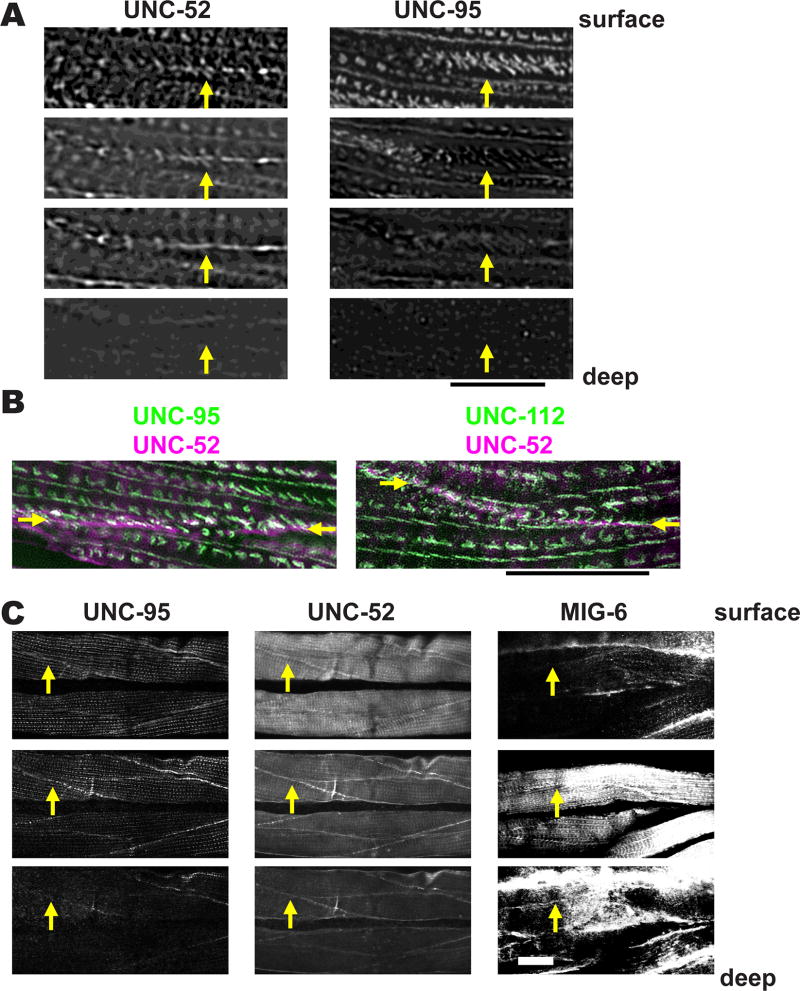
Figure 8. Muscle cell boundaries include extra cellular matrix proteins.
(A) Worms were co-immunostained with antibodies to UNC-95 and the ECM component UNC-52 (perlecan). Images were obtained by SIM, and each image was taken at 0.4 µm intervals. The yellow arrows point to muscle cell boundaries. Scale bar represents 10 µm.
(B) SIM images of a portion of two adjacent body wall muscle cells co-stained with either antibodies to UNC-52 and UNC-95 (left), or UNC-52 and UNC-112 (right). Arrows mark the muscle cell boundary. Note that most of the ECM protein UNC-52 lies between the zipper-like structures labeled with either UNC-95 or UNC-112. Scale bar, 10 µm.
(C) UNC-52 (perlecan) and MIG-6 (papilin, lacunin) localize at muscle cell boundaries both at “surface” (close to membrane adjacent to hypodermis) and “inside” (away from this surface) focal planes. Animals were immunostained with antibodies to UNC-95, UNC-52 and MIG-6, and images were obtained by confocal microscopy, and show three consecutive 1.0 µm optical sections. The yellow arrows point to muscle cell boundaries. White bar represents 10 µm.

