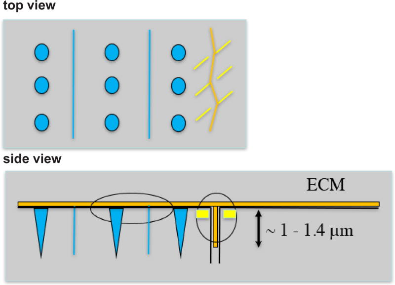
Figure 9. Drawing depicting ECM (orange), M-lines and dense bodies (blue) and attachment plaques at a muscle cell boundary (yellow).
The ECM lies at the base of the cells, and extends between cells for a distance of approximately 1–1.4 µm, based on Figure 8A. The attachment plaques do not extend as deeply as ECM between the muscle cells.

