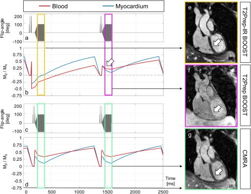
Figure 2.

Simulated magnetization of the proposed BOOST sequence (a, b) and a more conventional and dedicated CMRA sequence (c, d) after steady state has been reached. The expected longitudinal magnetization (Mz/M0) of myocardium (blue line) and blood (red line) is shown (b, d). With BOOST, the absolute signal ratio between blood and myocardium during data acquisition is optimized for odd heartbeats (ranging from 2.66 to 2.64, yellow rectangle). A strong contrast between the two tissues can be observed in the corresponding reconstructed T2Prep‐IR BOOST volume (e, arrow). Conversely, the absolute signal ratio between blood and myocardium is reduced in correspondence to the acquisition of even heartbeats (ranging from 1.35 to 2.11, purple rectangle). Specifically, such reduced signal ratio can be observed at the beginning of imaging data collection (arrow in b) and gets particularly emphasized with centric reordering acquisitions. As a consequence, reduced tissue contrast can be observed for the corresponding T2Prep BOOST reconstruction (f, arrow). Because T2Prep BOOST is used as the reference image in the process of PSIR reconstruction, reduced tissue contrast is indeed desired for adequate surface coil normalization and contrast of the resulting PSIR image. Conversely, and for the more conventional CMRA acquisition, the absolute blood to myocardium ratio ranges from 1.74 to 3.2 during data acquisition (green rectangles). Centric reordering acquisitions emphasize the improved blood to myocardium contrast with T2Prep‐IR BOOST in comparison to the dedicated CMRA. This can be appreciated in the corresponding image reconstructions (e, g, arrows). BOOST, Bright‐blood and black‐blOOd phase SensiTive; CMRA, conventional coronary MR angiography; T2Prep, T2 prepared.
