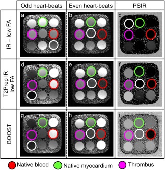
Figure 3.

Data acquisition in a standardized T1/T2 phantom using three different sequences: the PSIR sequence described in 22, alternating the acquisition of a T1‐weighted volume with the acquisition of a reference image acquired at low flip angle and without preparatory pulses (first row); the T2Prep‐IR PSIR sequence described in 23 that alternates the acquisition of a volume preceded by a T2Prep‐IR module with a reference acquisition performed at low FA angle and without preparatory pulses (second row); and the proposed BOOST sequence illustrated in Figure 1 (third row). Compartments of interest are indicated: blood (red circle), myocardium (green circle), and thrombus (purple circle). For all the images, SNRblood as well as CNRblood‐myo were computed with respect to a low‐signal compartment (white circle). With all the investigated sequences, the images acquired at odd heartbeats exhibit high SNRblood (a, d, g) and high CNRblood‐myo. Due to the relatively short T1 of the vial mimicking the coronary thrombus (purple circle), its signal appears suppressed in the images acquired at odd heartbeats with all the investigated sequences. The images (b) and (e), acquired at even heartbeats, show low SNR because data acquisition is performed at low FAs. Differently, higher SNRblood and CNRblood‐myo were observed with the proposed BOOST approach because data acquisition was performed with a 90‐degree FA. The PSIR reconstruction provided in (c) shows signal suppression for all the compartments of interest; differently, in the PSIR‐like reconstructions displayed in (f) and (i), a black‐blood effect is obtained, with remarkable depiction of the vial mimicking the thrombus. BOOST, Bright‐blood and black‐blOOd phase SensiTive; FA, flip angle; IR, inversion recovery; myo, myocardium; PSIR, phase sensitive inversion recovery; T2Prep, T2 prepared.
