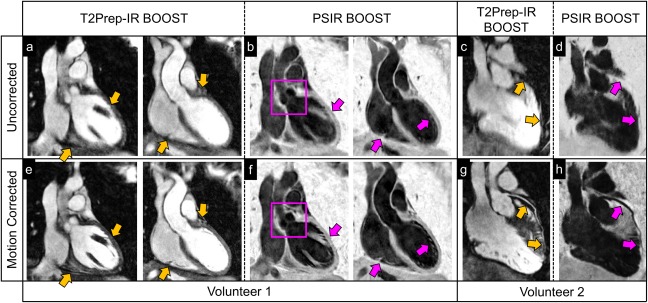
Figure 5.

Image quality improvement after motion correction in two healthy volunteers with the proposed BOOST framework. Bright‐blood T2Prep‐IR BOOST and black‐blood PSIR BOOST images are shown before (first row) and after (second row) motion correction in acquired coronal orientation (volunteer 1) and reformatted plane (volunteer 2). Yellow arrows show visible improvement in image quality at the level of the myocardium and of the interface between the heart and the liver for the motion‐corrected T2Prep‐IR BOOST dataset (a versus e). Similarly, a sharper depiction of anatomical structures (purple arrows), including the aortic valve (purple square), can be appreciated in the motion‐corrected black‐blood PSIR BOOST images (f) when compared to the uncorrected ones (b). For the second volunteer, motion correction successfully recovers a sharp visualization of the left anterior descendent coronary artery (LAD) for both the bright‐blood T2Prep‐IR BOOST dataset (c and g, yellow arrows) and the black‐blood PSIR BOOST dataset (d and h, purple arrows). BOOST, Bright‐blood and black‐blOOd phase SensiTive; IR, inversion recovery; PSIR, phase‐sensitive inversion recovery; T2Prep, T2 prepared.
