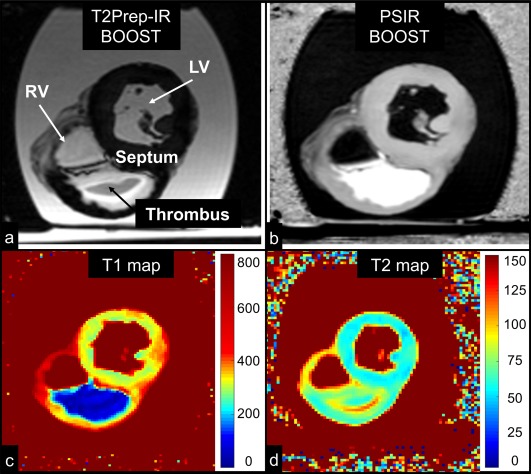
Figure 8.

MRI images obtained in the ex vivo pig heart. All the images depict a short‐axis view at the midventricular level. Images acquired with the proposed BOOST sequence are reported in (a) (bright‐blood T2Prep‐IR dataset) and in (b) (black‐blood PSIR‐like reconstruction). RV, LV, thrombus, and interventricular septum are indicated. The black‐blood reconstruction (b) clearly enhances the signal from the thrombus when compared to the bright‐blood dataset (a). Furthermore, 2D T1 (c) and T2 (d) mapping sequences were acquired. The ex vivo thrombus is characterized by a relatively short T1 and T2. BOOST, Bright‐blood and black‐blOOd phase SensiTive; IR, inversion recovery; LV, left ventricular cavity; PSIR, phase‐sensitive inversion recovery; RV, right ventricular cavity; T2Prep, T2 prepared.
