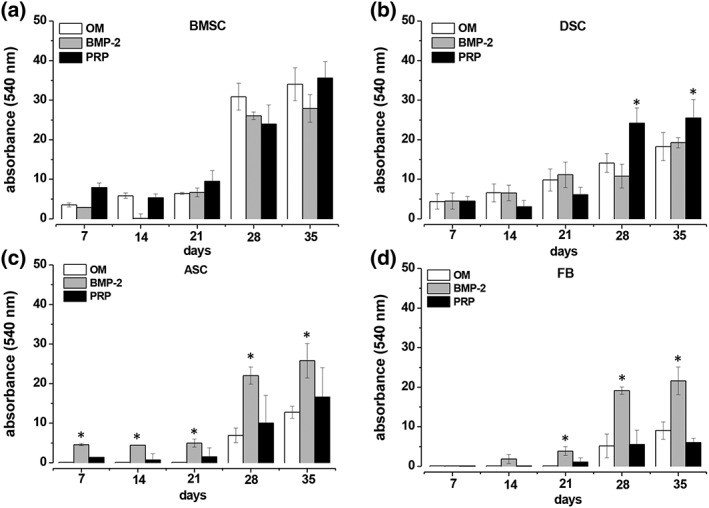
Figure 5.

Quantitative evaluation of the impact of BMP‐2 and platelet‐rich plasma (PRP) on osteogenic‐differentiated mesenchymal stromal cells (MSCs) obtained from bone marrow (BMSCs), adipose tissue (ASCs) and fibroblasts (FBs). BMSC (A), DSC (B), ASC (C) and FB (D) cultures were osteogenic differentiated using the standard osteogenic differentiation medium (OM). Additionally, OM (white bars) was supplemented with BMP 2 (grey bars; 450 ng/ml) or with PRP (black bars; 1%). At the time points indicated, the calcification of the extracellular matrix was quantified by staining with alizarin red S, extraction of the dye by cetylpyridiniumchlorid and photospectral analysis at 540 nm. Values in (A) and (D) represent the mean ± SD of four individual experiments, and the mean ± SD of six individual experiments in (B) and (C). * P < 0.05 as compared with the other values of the respective time points
