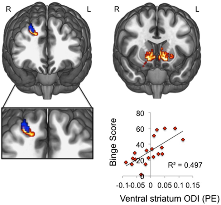
Figure 1.

Lower dorsolateral prefrontal cortex but higher ventral striatal orientation dispersion in binge drinkers. Left: clusters illustrate regions of significantly higher white matter neurite density (red) and lower grey matter orientation dispersion index (ODI, blue) in dlpfc of binge drinkers compared with healthy volunteers (displayed at p < 0.001 uncorrected threshold for visualization purposes). Right: small volume corrected family‐wise error p < 0.05 analyses revealed that binge drinkers had reduced ODI in ventral striatum compared with matched controls. Left sided peak ventral striatal ODI (peak coordinates, xyz = −6 12 0) positively correlated with binge score in binge drinkers. Images are displayed on a standard MNI152 template; R, right; L, left
