
Figure 4.
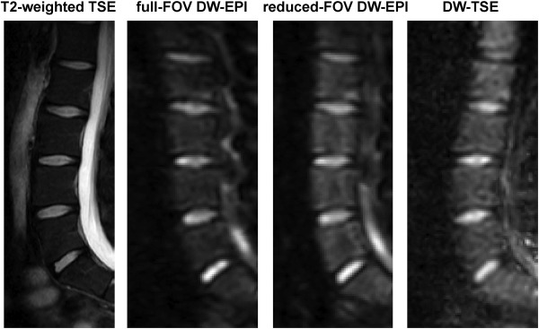
Diffusion‐weighted imaging of the spine in a 28‐year‐old male subject: T2‐weighted TSE for anatomical reference, full‐FOV DW single‐shot EPI (b = 800 s/mm2), reduced‐FOV DW single‐shot EPI (b = 800 s/mm2), single‐shot DW TSE (b = 800 s/mm2). Full‐FOV single‐shot EPI shows significant geometric distortions, which are reduced in reduced‐FOV single‐shot EPI and minimized in DW‐TSE. DW‐TSE shows increased blurring.