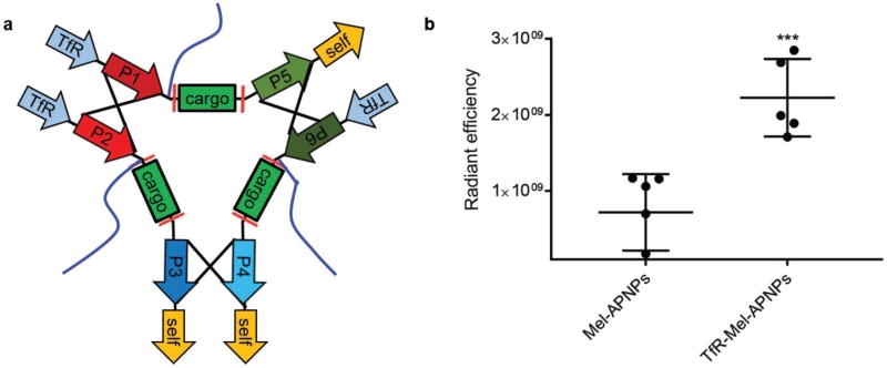
Figure 4.
TfR-Mel-APNPs for targeted delivery to tumors. a) Schematic of TfR-Mel-APNPs. b) In vivo quantitative distribution of Mel-APNPs with and without target ligand RTIGPSV. When tumor volume reached ≈200 mm3, mice were grouped and received treatment of AF750-labeled APNPs. 24 h later, mice were euthanized. The tumors were harvested and subjected to IVIS imaging. Fluorescence intensity in the ischemic region was determined using Living Image 3.0.

