Figure 1.

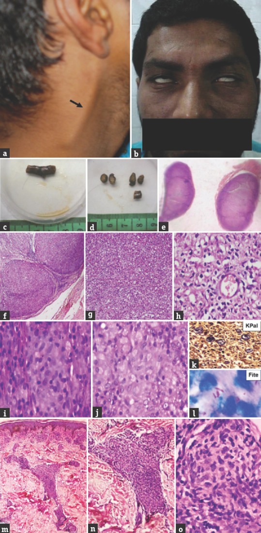
Case 1 mid-borderline – (a) Thickened right greater auricular nerve; (b) bilateral lower motor neuron facial palsy exhibiting lagophthalmos; (c and d) gross of thickened right greater auricular nerve; (e) whole-mount view of transverse section of thickened nerve; (f) enlarged fascicles (×40, H and E); (g) fascicles completely effaced by inflammatory cells (×100, H and E); (h) mononuclear cells, foam cells, and histiocytes (×200, H and E); (i and j) poorly formed granuloma (×400, H and E); (k) few remnant myelinated nerve fibers (×400, KPal); (l) lepra bacilli (OIF, Fite); (m) (×40, H and E), (n) (×100, H and E), (o) (×400, H and E); well-defined epithelioid granuloma in papillary and reticular dermis
