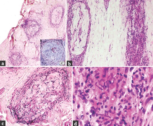
Figure 2.

Case 3 borderline tuberculoid – (a) Distinct inflammation in endo- and perineurium, marked endoneurial fibrosis (inset, Masson trichrome) and severe loss of myelinated nerve fibers (×40, H and E); (b) thickened perineurium and epineurial perivascular inflammation (×100, H and E); (c) thickened perineurium and endoneurial inflammation (×100, H and E); (d) epithelioid granuloma (×400, H and E)
