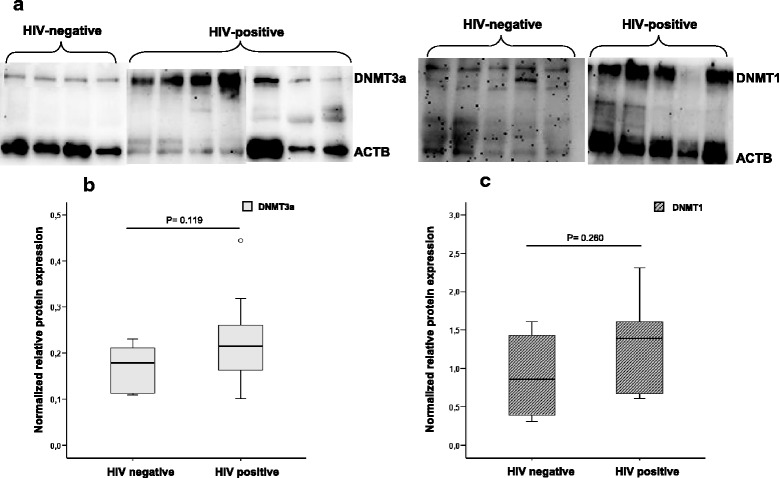
Fig. 3.

Western blot analysis. a Cropped gels/blots obtained by each protein evaluation. b, c Quantification of the expression of tested proteins and differences between HIV and non-HIV infected individuals: b DNMT3a and c DNMT1. The optical density of each sample was measured and normalized using an Actin run on the same gel. The data are expressed as relative expression (ratio DNMT/actin). Horizontal bars indicate median values, boxes indicate interquartile range (IQR), and p values for each protein are indicated
