Figure 3.

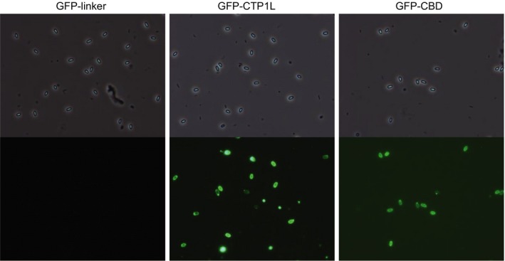
Fluorescent labelling of C. beijerinckii INIA 77 spores. Pictures were taken with phase contrast (top) and green fluorescence (bottom) at a magnification of ×1000.

Fluorescent labelling of C. beijerinckii INIA 77 spores. Pictures were taken with phase contrast (top) and green fluorescence (bottom) at a magnification of ×1000.