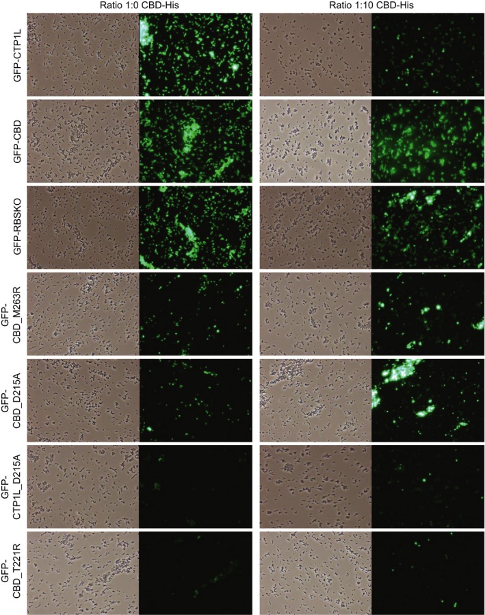
Figure 6.

Fluorescence microscopy images of binding of wild type and mutant GFP‐endolysins to C. tyrobutyricum NCIMB 9582 with (right column) and without (left column) addition of free CBD‐His. Pictures were taken with phase contrast and green fluorescence at a magnification of ×400.
