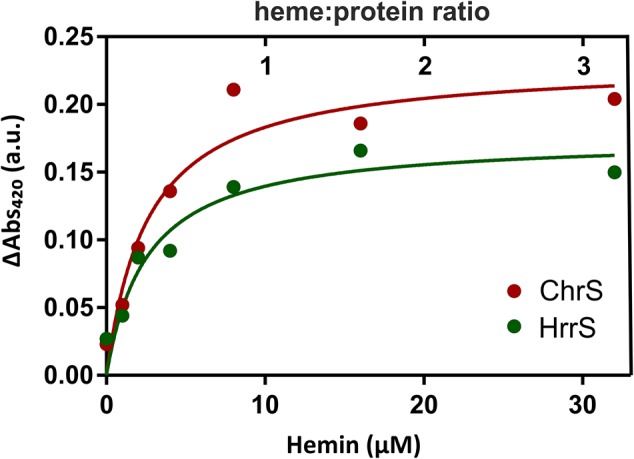
FIGURE 3.

UV-Vis analysis of the heme-binding properties of HrrS and ChrS. For hemin-binding assays, different amounts of hemin were titrated to 10 μM purified HrrS or ChrS to a final concentration of 0, 2, 4, 8, 16, and 32 μM. The mixture was incubated for 5 min at RT and then analyzed by UV-visual spectroscopy. The resulting absorption was referenced against the absorption of buffer containing only DDM micelles but no protein. The graph shows the maxima of the soret peaks at 420 nm.
