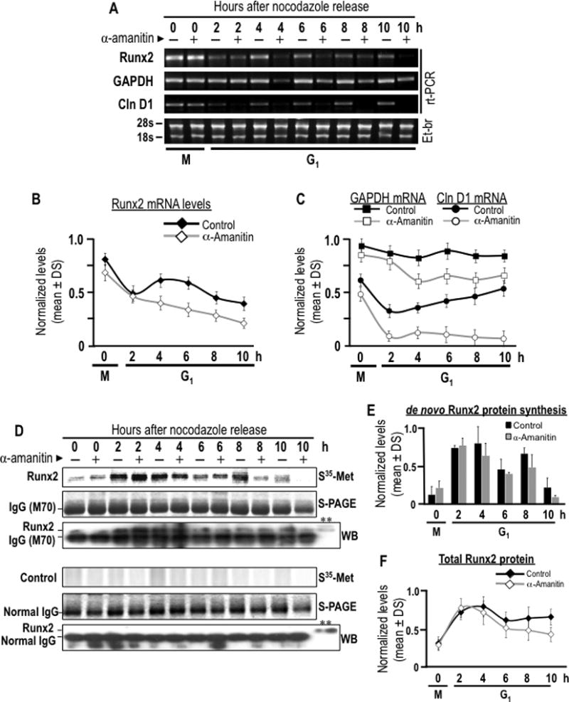
Fig. 7. Post-mitotic inheritance of Runx2 mRNA supports de novo Runx2 protein synthesis during early G1 phase.

The contribution of post-mitotically inherited Runx2 mRNA to Runx2 protein synthesis during G1 phase was assessed by combining mitotic cell synchronization with metabolic labeling. (A) MC3T3-E1 cells were synchronized with nocodazole to generate a mitotic block. Mitotic cells were pre-treated with or without α-amanitin for 2 h and harvested by gentle agitation (0 h) and then released through mitosis into G1 by washing and the addition of fresh culture medium supplemented with (+) or without (−) α-amanitin. Cells were harvested at selected time course during the M/G1 phase transition (0, 2, 4, 6, and 10 h). Runx2, cyclin D1 and Gapdh mRNA levels were analyzed by RT-PCR to evaluated α-amanitin effect on RNA polymerase II inhibition and transcripts stability. (B and C) Runx2, cyclin D1 and Gapdh mRNA levels showed in panel A are graphically represented. Significant statistically differences between the control and the treated group: (B) Runx2, for 4, 6, 8 and 10 h (P = 0.028, 0.013, 0.047 and 0.031, respectively), (C) cyclin D1, for 2 to 10 h (P = 0.04, 0.032, 0.018, 0.009 and 0.003, respectively), and Gapdh, from 4 h (P = 0.026, 0.021, 0.017 and 0.036, respectively). Cells were metabolically labeled by 2 h pulse with [35S]-methionine and harvested at selected time course during the M/G1 phase transition (0, 2, 4, 6, and 10 h). (D) Cells pulse-labeled with [35S]-methionine were subjected to immuno-precipitation using a Runx2 polyclonal antibody or non-specific IgG control. Immuno-precipitates of endogenous Runx2 were separated in by SDS-PAGE (S-PAGE) and de novo synthesis of Runx2 protein was assessed by autoradiographic (35S-Met-Arg) analysis. Immuno-precipitations were analyzed by western blot analysis with a Runx2 monoclonal antibody or non-specific IgG control. A representative input was included to validate immuno-reactive Runx2 bands (**). Note that Runx2 is observed immediately above the immunoglobulin heavy chain. The data shown is representative of three experiments with similar outcomes. (E and F) Bar and line graphs show autoradiographic and western blot signals for immuno-precipitated Runx2 showed in panel D, no statistical differences between the control group and treated.
