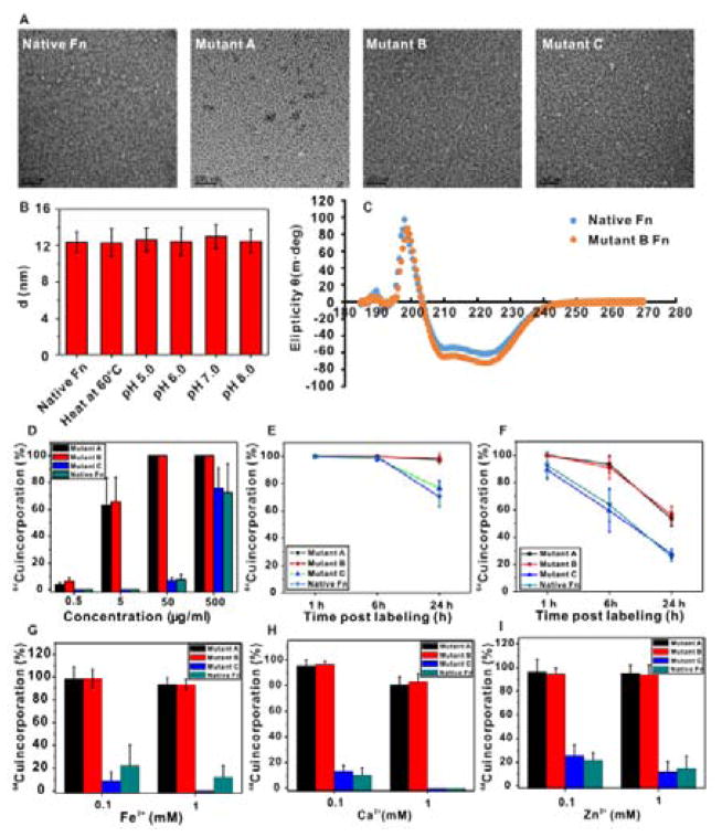
Figure 2.
(A) TEM images of ferritin variants with uranyl acetate negative staining. Scale bar=100 nm. (B) Dynamic light scattering study of the nanocage size of mutant B ferritin under heating (60 °C) and different pH values in buffers. (C) Circular dichroism spectra of native ferritin and mutant B ferritin in PBS (0.5 mg/mL). (D) Radioactive 64Cu incorporation ratio of ferritin variants under different concentrations. (E) 64Cu incorporation stability study of ferritin variants in mouse serum over 24 h. (F) 64Cu incorporation stability study of ferritin variants in the presence of strong metal chelator, EDTA, in mouse serum over 24 h. (G–I) Divalent metal ion competition with 64Cu in ferritin variants.

