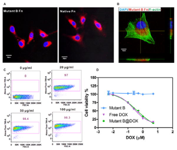
Figure 3.
(A) Cancer cell (U87MG) uptake of ferritin variants labeled with Cy5.5 fluorophore. Scale bar = 20 μm (B) Magnified confocal laser scanning imaging (CLSM) (Z-section) of intracellular uptake of mutant B ferritin after 2 h incubation. Scale bar = 10 μm. Blue: nucleus counterstained with DAPI; Red: Cy5.5 labeled mutant B ferritin; Green: F-actin stained with Alexa488 conjugated phalloidin. (C) Flow cytometry analysis of cellular uptake of mutant B ferritin at different concentrations. (D) MTT assay of mutant B ferritin with cancer cells (n = 6).

