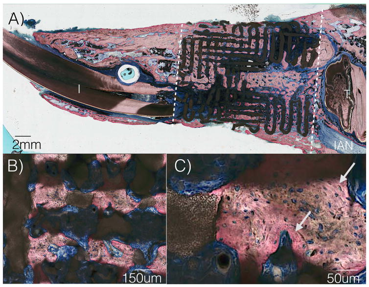
Figure 4.
(a) Sagittal histologic slice of scaffold in continuity with rabbit mandible (b) High magnification from 4a demonstrating porous ingrowth (c) Highly cellular and vascularized woven bone structure, as well as newer, organized lamellar bone formation depicted by arrows. (I=Incisor, T= tooth, IAN= inferior alveolar nerve)

