Figure 6.
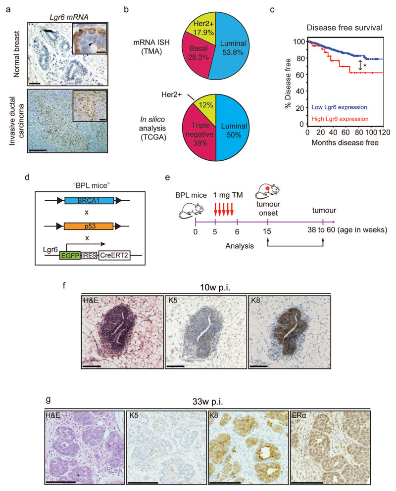
Lgr6+ cells are present in human breast carcinoma and are able to initiate tumours in mouse models of breast cancer. (a) In situ hybridisation (ISH) analysis of Lgr6 mRNA expression on human breast carcinoma tissue microarrays (TMA) compared to normal healthy breast tissue. Keratin 5 (K5) co-staining demonstrates the presence of basal and luminal Lgr6+ mammary gland cells (inset, upper panel). Scale bars: 100µm, 20µm (insets) (b) Prevalence of Lgr6 expression or up-regulation within the three categories of human breast tumours as determined by ISH on tissue microarrays (n = 550 tumours; upper pie chart) and in silico analysis of breast cancer gene expression data available from The Cancer Genome Atlas (TCGA; lower pie chart) (cBioportal, RNASeq, and ref59. (c) Disease free survival in n = 1093 patients with Lgr6 up-regulation compared to control patients (cBioportal, RNASeq, and ref 59). *P = 0.042 (log-rank test). (d) Description of the BPL mouse strain, derived from breeding of BRCA1f/f, p53f/f, and Lgr6-CreERT2 mice. (e) Treatment schedule of 5w-old BPL mice with tamoxifen and subsequent tumour analysis. (f) Immunohistochemistry of tumours from BPL mice 10 weeks post-injection with tamoxifen, stained with haematoxylin-eosin (HE) and probed for markers against basal (K5+) and luminal (K8+) cells. Scale bars: 100µm. (g) Immunohistochemistry of tumours from BPL mice 33 weeks post-injection stained with HE and probed for K5, K8 and ERα. Scale bars: 100 µm.

