
Figure 2.
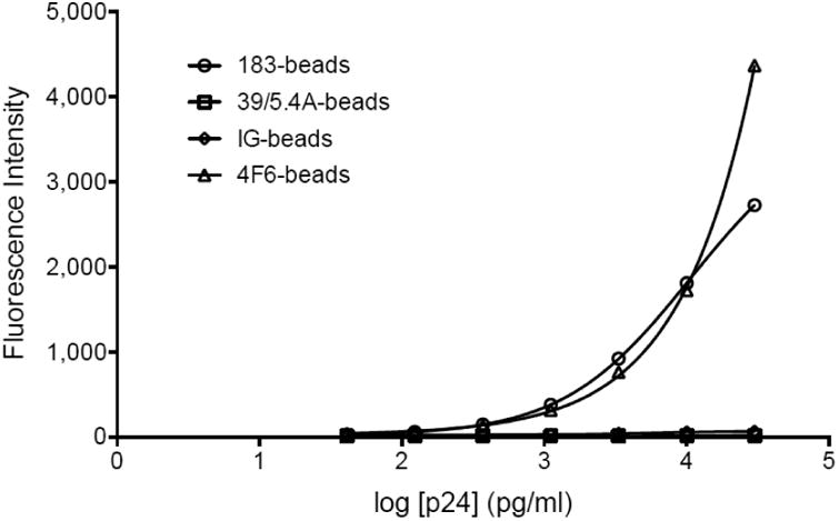
Validation of the functionality of the capture antibodies post coupling. The capture microspheres (183-beads, empty circles; 39/5.4A-beads, empty squares; IG-beads, empty diamonds and 4F6-beads, empty triangles) were mixed with serial dilutions of purified p24 protein, the binding was revealed by PE-labeled anti-p24 and read by Luminex. The graph represents standard curves fitted by a logistic 5P regression using Prism6.