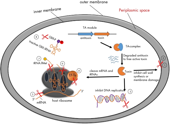
Figure 1.
The intracellular targets of TAloci. TA loci usually encode two genes: one is a stable toxin and the other one is an unstable antitoxin. The antitoxins sequester the toxins but are subjected to proteolytic degradation by cellular proteases (Lon or ClpXP) under stress condition. Consequently, free active toxins alter cellular processes including DNA replication, translation or cell-wall synthesis, which ultimately results in slow growth or the formation of highly drug-tolerant persisters. TAs examples for the cellular targets are given below. (1) Zeta toxin inhibits cell-wall synthesis by specific phosphorylation of peptidoglycan precursor UNAG. (2) TisB, HokB and GhoT: the products of TisB and HokB can decrease the level of membrane potential motive force (pmf) and ATP by inserting into cytoplasmic membrane, while protein GhoT can lyse cell membrane and change cell morphologies. (3) CcdB and ParE inhibit DNA replication by poison DNA gyrase. (4) Doc inhibits translation by phosphoralation of elongation factor Tu (EF-Tu). (5–7) MazF, RelB and VapC inhibit translation by cleavage of mRNAs like single-stranded mRNA, A-site on ribosome and initiator tRNAfMet, respectively. (8) HipA inhibits translation by phosphoration of GltX. tRNA:fMet indicates initiator tRNA at P site carried formyl methionine; ‘p’ indicates phosphorylation.

