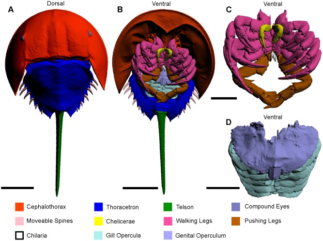
Fig 1. 3D reconstruction of the complete museum specimen (va.06) of Limulus polyphemus in dorsal and ventral aspect, as modelled from CT scanning.
(A) Dorsal view of entire exoskeleton. (B) Ventral view of entire exoskeleton. (C) Detail of cephalothoracic appendages. (D) Detail of thoracetronic appendages. The interactive 3D version of this file is available in the Supporting Information (S1 Fig). Scale bars 100 mm for A, B; 50 mm for C, D.

