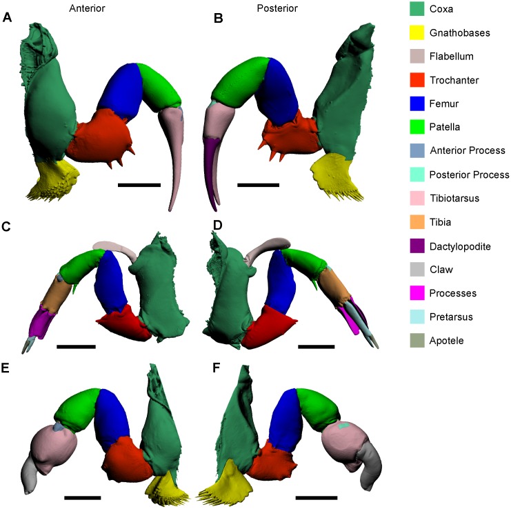
Fig 3. 3D reconstructions of the walking leg (A, B), pushing leg (C, D), and male pedipalp (E, F) as modelled from micro-CT scanning.
Reconstructions in the left column (A, C, E) are all anterior views and reconstructions in the right column (B, D, F) are all posterior views. These reconstructions should be studied in conjunction with the muscle reconstructions in Fig 4. The 3D versions of these files are available in the Supporting Information (S3, S4 and S5 Figs). Scale bars 15 mm for A, B, E, F; 20 mm for C, D.

