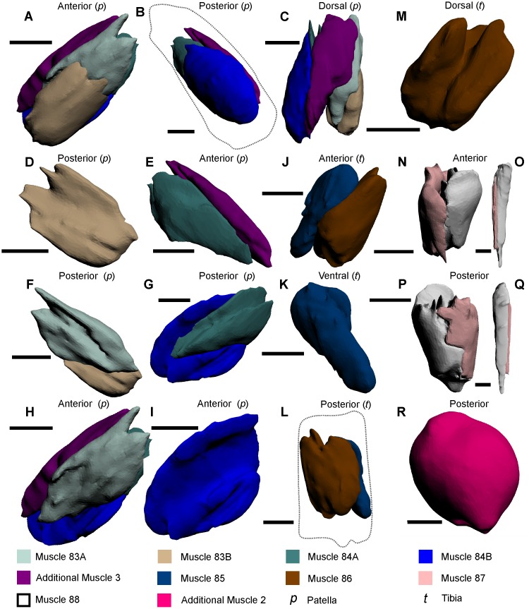
Fig 8. The muscles within the patella, tibia and pretarsus of the pushing leg, and the walking leg and male pedipalp tibiotarsus muscles, as modelled from iodine staining and micro-CT–scanning.
(A–C) The five muscles within the patella of the pushing leg in three views to illustrate the overall form of the muscles. (D–I) Each of the five muscles in anterior and posterior views to accurately display the main features and shapes thereof and, where possible, their position relative to adjacent muscles. (J–M) The two muscles within the tibia of the pushing leg. (N, P) The two muscles within the tibiotarsus of the walking leg. (O, Q) The two muscles within the pretarsus of the pushing leg. (R) The newly described muscle within the tibiotarsus of the male pedipalp; due to the subspherical nature thereof, the anterior perspective was not included. Muscle reconstructions A–M, O and P were selected from the pushing leg 3D PDF (S5 Fig). The muscle reconstructions of N and P were selected from the walking leg 3D PDF (S3 Fig). The muscle reconstruction of R was selected from the male pedipalp (S4 Fig). The dotted line in B outlines the pushing leg patella. The dotted line in L outlines the tibia. Scale bars 5 mm for A–N, R; 4 mm for P; 3 mm for O, Q.

