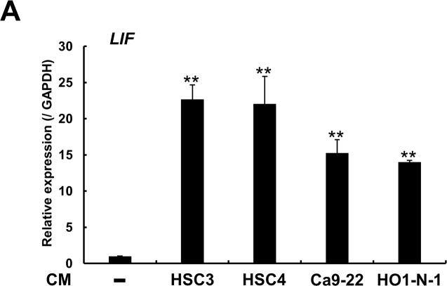
Fig 2. OSCC cell lines stimulate LIF expression in NHDFs.
CM derived from 4 OSCC cell lines were separately added to NHDFs, and increased LIF expression was confirmed through quantitative real-time PCR analysis after 48 h of culture. The experiment was performed in triplicate in 24-well plates. Representative graph from 3 independent experiments is shown. Data represent means ± SEM. Multiple comparisons were performed by using one-way ANOVA with Dunnett’s method. **p < 0.01 compared with control NHDFs.

