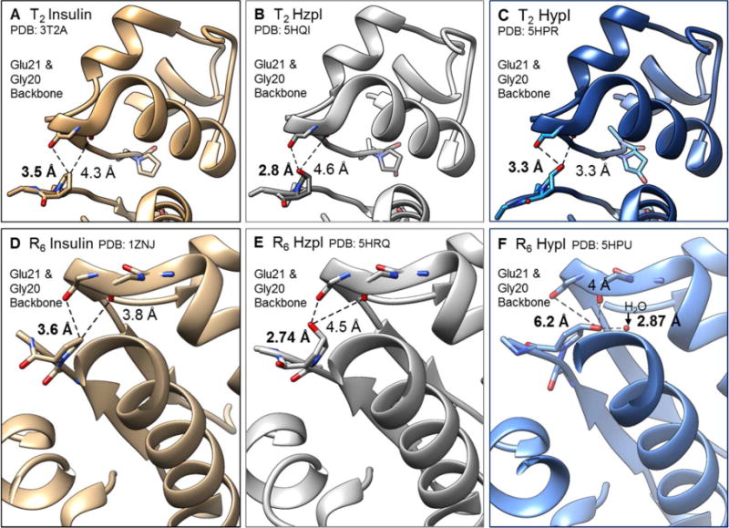
Figure. 3. Crystal structures of HzpI and HypI.

In tan (left), wt insulins from PDB (3T2A, 1ZNJ) highlighting the distance between the carbon atom at the 4th position of ProB28 and its closest neighbors, backbone carbonyl oxygen atoms of GlyB20’ and GluB21 in the T2 dimer (A) and R6 hexamer (D) forms. In grey (middle), HzpI in the T2 dimer (B) and R6 hexamer (E) forms. In blue (right), HypI in the T2 dimer (C) and R6 hexamer (F) forms.
