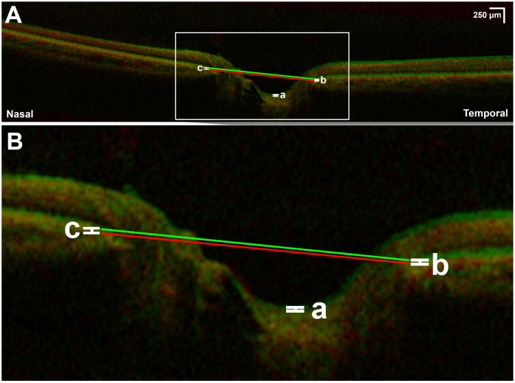
Fig. 3.
A) Superimposed optical coherence tomography of left eye demonstrating effect of 30° abduction (green) compared to central gaze (red). B) Magnification of rectangular area indicated in Fig 3A. There is minimal anterior displacement of the optic cup (a), temporal peripapillary retinal pigment epithelium (b), and nasal peripapillary retinal pigment epithelium (c). Lines defining optic nerve head angle in central gaze (red line) and abduction (green line) are parallel, indicating absence of angle change with 30° abduction.

