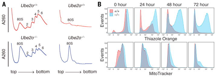
Fig. 2. Ube2o−/− reticulocytes have elevated levels of ribosomes.
(A) Polysome profiles were generated by fractionating Ube2o−/− and WT reticulocyte lysates on 20–50% sucrose gradients. The X-axis represents position in the gradient, the Y-axis OD260. Left, freshly-isolated Ube2o−/− reticulocytes showed an 80S monosome peak that is elevated in comparison to WT. Right, as above but after 31 hours of ex vivo differentiation.
(B) Flow cytometry analysis after ex vivo differentiation. WT CD71+ reticulocytes showed progressive loss of both MitoTracker Deep Red and thiazole orange staining. Ube2o−/− CD71+ reticulocytes retained thiazole orange staining after 72 hours of ex vivo differentiation (upper panels), with mitochondrial elimination unaffected (lower panels).

