
Fig. 5. Characteristics of the CEMs obtained from gene 20 spacers.
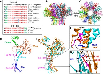
(A) List of CEM sequences obtained from gene 20 spacers. The spacer sequence and PAM are shown in red and green, respectively, and the mutated nucleotides are shown in blue (bold) and underlined. The WT sequences are shown at the top of each alignment. (B to G) Structural analysis of the CEMs of the portal protein gp20. Side (B) and top (C) views of the structure of the dodecameric gp20 portal assembly with each subunit shown in a different color. Single (D) and two (E) subunits of gp20 showing (i) the critical salt bridge between the D361 residue of one subunit and R275 residue of an adjacent subunit (circled) (F) and (ii) the functionally important residues of the clip domain, E332 and D333, of one subunit forming a salt bridge with R311 of an adjacent subunit (circled) (G). The regions corresponding to the protospacer and PAM sequences of spacers 20-1070 (D, E, and F) and 20-995 (D, E, and G) are shown in magenta. The positions of the mutated residues of the CEM phages corresponding to spacers 20-1070 (W364) (F) and 20-995 (T331 and L336) (G) are shown with arrows.