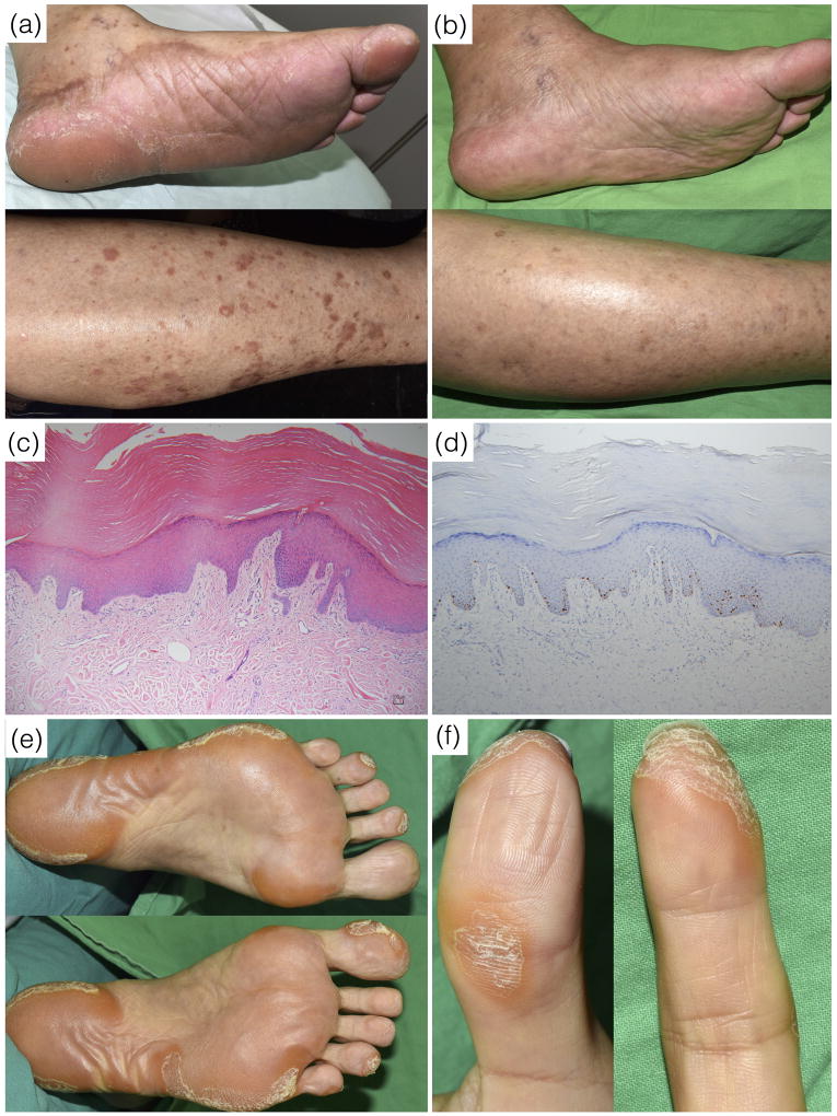
Fig. 1.
Patient 1. (a) About one month after the initiation of olmutinib therapy, brownish asymptomatic dispersed hyperkeratotic patches and plaques developed on the bilateral soles of the feet and shins. (b) The lesions resolved spontaneously at 1 month after discontinuation of olmutinib. (c) The pathology study showed acanthosis, hyperkeratosis and papillary dermal elongation with minimal dermal inflammation (hematoxylin and eosin stain, 100X). (d) The immunohistochemistry study showed Ki-67 positive cells confined to the basal and suprabasal layers (Ki-67 stain, 100X). Patient 2. (e) Asymptomatic, focal hyperkeratotic plaques mainly on the pressure points of the soles and (f) tips of the toes and fingers, but sparing the nails.

