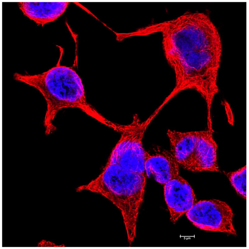
Figure 3.

Fluorescence microscopy images of basal cell carcinoma cell line showing the microtubule cytoskeleton and the nucleus. The cells are UW-BCC1 cells (58), grown on glass coverslips and imaged. Microtubules (red) were imaged by immunostaining with monoclonal anti-alpha tubulin antibody and the DNA in the nucleus (blue) was revealed by Hoechst stain.
Scale bar is 20 μm.
