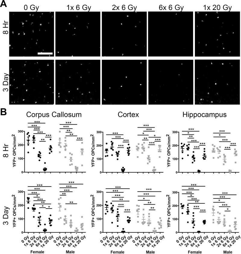
Figure 2. Fractionation enhances radiation-induced loss of oligodendrocyte progenitor cells.
A. Representative images of cortical yellow fluorescent protein (YFP) expressing cells 8 hours and 3 days after the final exposure to 0, 1× 6, 2× 6, 6× 6 or 1× 20 Gy irradiation. Scale bar = 200 µm. B. At both 8 hours and 3 days, the number of YFP+ OPCs was reduced by radiation in the corpus callosum, cortex, and hippocampus (p < 0.0001 for all). At 8 hours, an effect of sex was observed in the corpus callosum (p = 0.0250). At 3 days, an effect of sex was observed in the corpus callosum (p = 0.0002), cortex (p = 0.0006), and hippocampus (p = 0.0032). n = 5–8 animals/group, 2-way ANOVA with Bonferroni’s multiple comparisons test comparing all conditions. Graphs represent mean ± SEM. *p<0.05, **p<0.01, ***p<0.001. Graphs show statistically significant differences within sexes only. Significance was also found in the 1× 6 Gy IR female versus male groups at 3 days in the corpus callosum (p < 0.05) and cortex (p < 0.05) with Bonferroni’s post-test.

