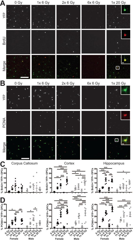
Figure 4. Proliferation of oligodendrocyte progenitor cells is enhanced by radiation exposure.
Three days after the final dose of radiation, proliferating OPCs were labeled by intraperitoneal injection of 5-bromo-2-deoxyuridine (BrdU) 2 hours prior to sacrifice. Representative images show enhanced BrdU (A) and PCNA (B) expression in cortical yellow fluorescent protein (YFP)-positive OPCs after radiation exposure. Scale bars = 200 µm. Inset scale bars = 10 µm. C. Radiation significantly increased the proportion of OPCs in S-phase of the cell cycle in the corpus callosum (p = 0.0099), cortex (p < 0.0001), and hippocampus (p < 0.0001). D. The PCNA labeled fraction of OPCs was affected by radiation dose in all areas examined (p < 0.0001 for each). n = 5–8/group, 2-way ANOVA with Bonferroni’s multiple comparisons test comparing all conditions. Graphs represent mean ± SEM. *p<0.05, **p<0.01, ***p<0.001.

