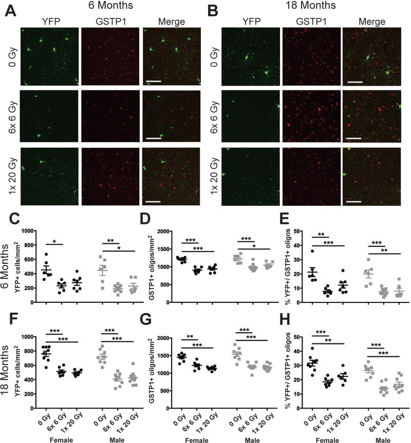
Figure 7. Reduced maturation of oligodendrocyte progenitor cells correlates with decreased numbers of mature oligodendrocytes at 6 and 18 months after radiation exposure.
A., B. Representative images of cortical YFP and GSTP1 expressing cells at 6 and 18 months from radiation. Scale bar = 50 µm. C. At 6 months, an effect of radiation dose (p < 0.0001) on YFP+ cells was observed. D. Both radiation dose (p < 0.0001) and sex (p = 0.0227) affected GSTP1 cell numbers at 6 months. E. The proportion of GSTP1+ oligodendrocytes that expressed YFP was reduced by radiation dose at 6 months (p < 0.0001). F. At 18 months, an effect of radiation dose (p < 0.0001) and sex (p = 0.0064) on YFP+ cells was observed. G. Radiation dose (p < 0.0001) affected the number of GSTP1 cells at 18 months. H. Radiation dose (p < 0.0001) and sex (p = 0.0003) affected the proportion of GSTP1+ oligodendrocytes that expressed YFP. n = 6–9/group. 2-way ANOVA with Bonferroni’s multiple comparisons test comparing all groups. Graphs represent mean ± SEM. *: p<0.05, **: p<0.01, ***: p<0.001.

