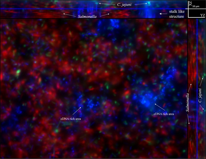
FIG 8.
Spatial distribution of C. jejuni cells, Salmonella cells, and extracellular DNA (eDNA) within a Campylobacter-Salmonella dual-species biofilm formed in the microfluidic platform. Fluorescence microscopy was applied to determine the spatial distribution of eDNA and bacterial cells in a dual-species Campylobacter-Salmonella biofilm. After a 3-day cultivation, 30 nM DAPI solution was injected into the microfluidic device to stain eDNA in the biofilm. Images were collected at multiple channels: 405 nm (blue for DAPI signal), 488 nm (green for GFP signal), and 543 nm (red for RFP signal). Within this Campylobacter-Salmonella dual-species biofilm, C. jejuni cells were mainly located at the bottom whereas Salmonella cells were mainly located at the top layer. The eDNA mainly assembled and formed several eDNA-rich areas and maintained a spatial distance between C. jejuni and Salmonella in the biofilm.

