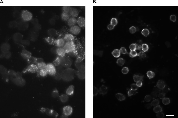
FIG 2.
Detection of enterovirus VP1 protein by immunofluorescence staining on HEp-2 cells. (A) Positive (infected) cells show punctate fluorescence, mainly in the cytoplasmic region. Samples are from the body of the dam. (B) Positive (infected) cells show fluorescence at the cellular periphery (membranous ring). Samples are from the beaches of the dam. Scale bar, 20 μm.

