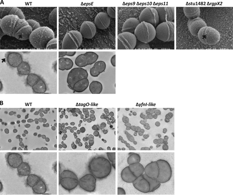
FIG 1.
Scanning electron microscopy (SEM) and transmission electron microscopy (TEM) observations of WT LMG 18311 and mutants. (A) SEM observations (original magnification, ×50,000) (upper panels) and TEM observations (original magnification, ×10,000) (lower panels) for the indicated strains. Black arrows indicate EPS. (B) TEM observations (original magnification, ×2,500 [upper panels] and ×10,000 [lower panels]) for the epsE mutant compared to the WT.

