Figure 1.
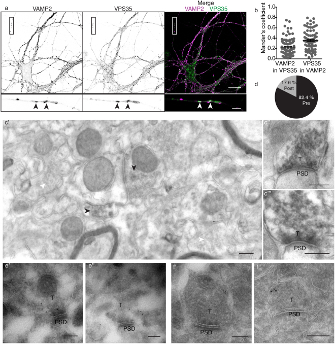
VPS35 is present in presynaptic terminals. (a) Representative confocal microscopy images of hippocampal neurons immunolabeled for VAMP2/synaptobrevin and VPS35. Arrowheads indicate co-localization between VAMP2/synaptobrevin and VPS35 puncta. Scale bar of the neuron image = 20 μm, scale bar of the zoomed neurite = 3 μm. (b) Mander’s coefficients for the co-localization between VAMP2/synaptobrevin and VPS35 in neurites (n = 78 fields of view, N = 3 animals). (c) Representative electron micrographs of hippocampal synapses from 3 independent wild-type mice. Each of the micrographs correspond to a different animal (N = 3 animals). Scale bar = 200 nm. (c’) In black arrowheads indicate the synaptic vesicle cloud of DAB positive presynaptic terminals and in white arrowheads DAB negative presynaptic terminals. (c”,c”’). Zoom in of DAB positive presynaptic terminals. (d) Percentage of synapses with VPS35 immunoreactivity in the presynaptic site (82.4% Pre) and postsynaptic site (17.6% Post). (e’,e”) Immunoelectron micrographs of presynaptic terminals stained with a rabbit antibody against VPS35 labelled with Protein A-10nm gold conjugate. The images are representative of two independent experiments (N = 2 animals). Scale bar = 200 nm (f’,f”) Immunoelectron micrographs of presynaptic terminals stained with a goat antibody against VPS35 labelled with a secondary antibody rabbit anti-goat and Protein A-10nm gold conjugate. The images are representative of two independent experiments (N = 2 animals). Scale bar = 200 nm. ‘PSD’ indicates postsynaptic side and ‘T’ the presynaptic terminal.

