Figure 3.
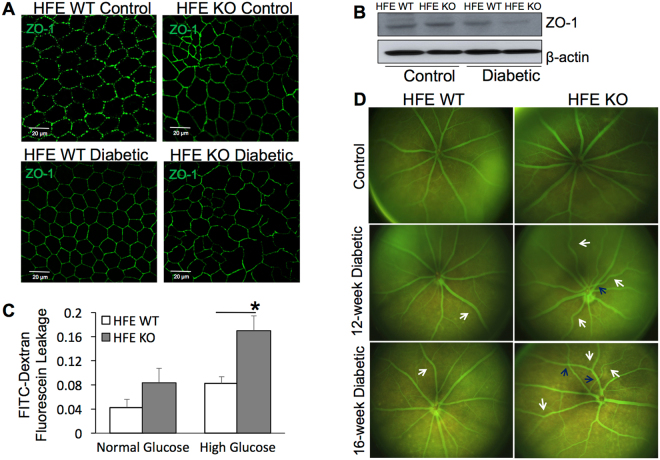
Altered outer and inner retinal barrier function in diabetic HFE KO mice. (A) Tight junction protein Zonula occludin-1 (ZO-1) immunofluorescence in retinal pigment epithelium (RPE) flat mounts prepared from non-diabetic control and 16-week post-diabetic HFE WT and KO mice. (B) Western blot analysis of ZO-1 expression in protein extracted from the RPE of non-diabetic and diabetic HFE WT and KO mice. β-actin was used as a loading control. Blots cropped from different parts of the same gel or from different gels are separated by white space. (C) FITC-Dextran (40-kD) trans epithelial permeability assay performed on confluent monolayers of primary RPE cells established from HFE WT and HFE KO mouse eyes grown in normal glucose (5.5 mM) or high glucose (25 mM). Data are presented as mean ± S.E. *p < 0.01. (D) Fluorescein angiography and fundoscopic imaging of non-diabetic control vs diabetic HFE WT and KO mice.

