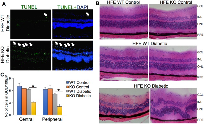
Figure 4.
Accelerated neuronal cell loss and morphological changes in retina of diabetic HFE KO mice. (A) TUNEL assay in the eye sections of diabetic HFE WT and KO mice. Green signals indicate apoptotic cells. (B) Hematoxylin and eosin stained retinal sections from non-diabetic HFE WT and KO mice (top panel) compared to diabetic HFE WT (middle panel) and diabetic HFE KO mice (bottom panel) (C) Quantification of number of cells in ganglion cell layer in the central and peripheral regions of retinal sections from non-diabetic and diabetic HFE WT and KO mice. *p < 0.01.

