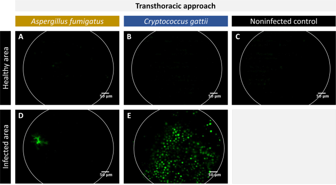
Figure 2.
In situ, transthoracic FCFM of lung infections. The images were acquired by placing the fibre-optic probe (S-1500) into direct contact with the lung surface after performing a bilateral thoracotomy. (A–C) Representative FCFM images of a healthy appearing area of an A. fumigatus (GFP-expressing) infected lung, a C. gattii (GFP-expressing) infected lung and a non-infected control lung. (D,E) Representative FCFM images of an infected area of an A. fumigatus infected lung and a C. gattii infected lung, showing the presence of fungal cells.

