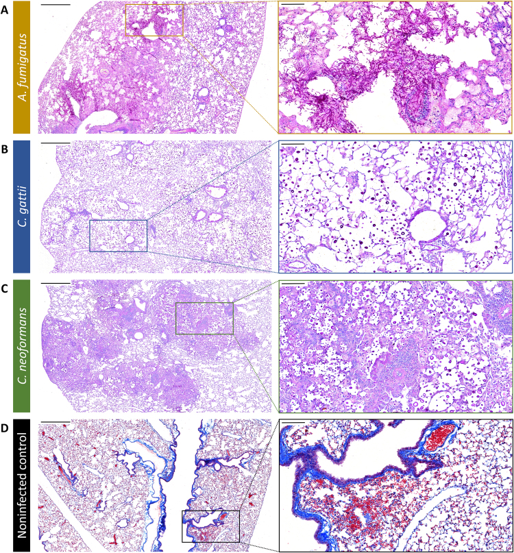
Figure 7.
Validation of the in vivo bronchoscopic FCFM results by histology. Following the last imaging time point, all animals were sacrificed and the lungs were isolated for histological analysis. (A–C) Representative light microscopy images (Periodic Acid-Schiff staining) showing an A. fumigatus, C. gattii or C. neoformans infected lung section, confirming infection in the imaged animals. (D) Representative light microscopy images from a non-infected control animal showing small haemorrhages within the lung tissue (Masson’s Trichrome staining). Scale bars measure 500 µm (left) or 100 µm (right).

