Fig. 1.
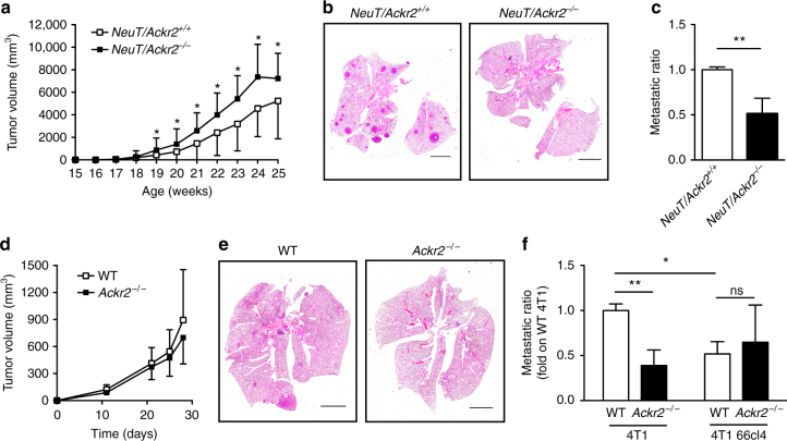
Ackr2−/− mice are protected from lung metastasis. a NeuT/Ackr2+/+ (white squares) and NeuT/Ackr2−/− (black squares) mice were evaluated for tumor growth calculated as described in the Materials and Methods section (n = 42 for NeuT/Ackr2+/+ and 23 for NeuT/Ackr2−/− mice). b Representative images of hematoxylin and eosin staining of NeuT/Ackr2+/+ and NeuT/Ackr2−/− lungs at 25 weeks of age. Magnification: ×10. Scale bar: 3 mm. c Metastatic ratio of NeuT/Ackr2+/+ (white column) and NeuT/Ackr2−/− (black column) mice, calculated as described in the Materials and Methods section (n = 26 and NeuT/Ackr2+/+ and 16 for NeuT/Ackr2−/− mice, respectively). d Tumor volume in WT (white symbols) and Ackr2−/− (black symbols) mice injected orthotopically with 4T1 cells (n = 14 for WT and 13 for Ackr2−/− mice). e Representative images of hematoxylin and eosin staining of WT and Ackr2−/− lungs at day 28 after 4T1 cell injection. Magnification: ×10. Scale bar: 3 mm. f Metastatic ratio of WT (white columns) and Ackr2−/− (black columns) mice at day 28 after orthotopic injection of 4T1 or 4T1 66cl4 cells (n = 14 for WT and 13 for Ackr2−/− mice for 4T1, 4 for both WT and Ackr2−/− mice for 4T1 66cl4). Data are represented as mean (SD). p value was generated using the unpaired t-test. *p < 0.05, **p < 0.01, ns = not statistically different

