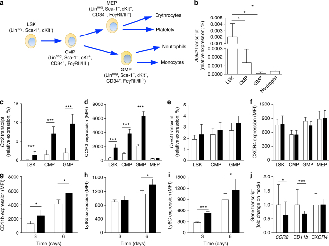
Fig. 6.
Ackr2 is expressed in HPCs and controls expression of CC chemokine receptors. a Schematic representation of murine hematopoietic cell differentiation. b qPCR analysis of Ackr2 expression on sorted HPCs and neutrophils taken from BM of WT mice (n = 7). c qPCR analysis of Ccr2 and e Cxcr4 expression on sorted HPCs taken from WT (white columns) and Ackr2−/− (black columns) mice. All qPCR data are relative to β-actin expression (n = 7 for both WT and Ackr2−/− mice). d MFI of CCR2 and f CXCR4 expression measured by FACS analysis in HPCs taken from WT (white columns) and Ackr2−/− (black columns) mice (n = 4 for both WT and Ackr2−/− mice). g MFI of CD11b, h Ly6G, and i Ly6C expression on FACS-sorted LSK cells taken from WT (white columns) and Ackr2−/− (black columns) cultured in vitro as described in Materials and Methods (n = 5 for WT and 7 for Ackr2−/− at 3 days, and 5 for WT and 6 for Ackr2−/− at 6 days). j qPCR analysis of CCR2, CD11b, and CXCR4 expression in HL-60 cells transfected with ACKR2 (black columns) or empty vector (white columns). qPCR data are relative to GAPDH expression and normalized on mock transfected cells (n = 9 for mock and 13 for ACKR2-transfected cells, three independent experiments). Data are represented as mean (SD). p value was generated using the unpaired t-test. *p < 0.05, ***p < 0.001

