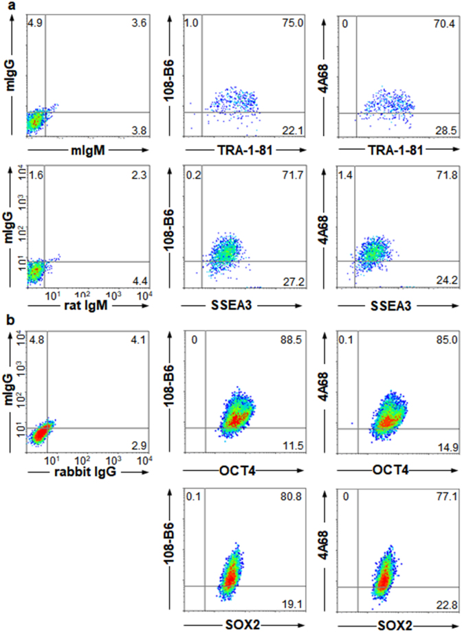
Figure 1.
Expression of 108-B6 and 4A68 antigens is localized to undifferentiated and pluripotent hPSCs. (a) Multi-color flow cytometric analysis of hPSCs. H9 hPSCs were stained with MAbs (108-B6 or 4A68) and PE-conjugated anti-mouse IgG. The cells were then stained with anti-TRA-1-81 or anti-SSEA3 antibodies followed by incubation with FITC-conjugated anti-mouse IgM or anti-rat IgM, respectively, before analysis. (b) Multi-color intracellular flow cytometric analysis of hPSCs. H9 hPSCs were stained with MAbs 108-B6 or 4A68 and PE-conjugated anti-mouse IgG. The cells were fixed with 2% PFA and incubated in PBA containing 0.5% saponin. The cells were then stained with anti-OCT4 or anti-SOX2 antibodies followed by incubation with Alexa 488-cojugated anti-rabbit IgG before analysis.

