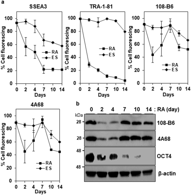
Figure 3.
Expression of 108-B6 and 4A68 antigens upon differentiation. (a) Flow cytometric analysis of the binding percentages of anti-TRA-1-81, anti-SSEA3, 108-B6 or 4A68 antibodies to undifferentiated H9 hPSCs (ES) or retinoic acid-treated H9 cells (RA). Undifferentiated H9 hPSCs were cultured in the presence of RA (10−5 M) for 14 days. ES or RA cells were incubated with anti-TRA-81, anti-SSEA3, 108-B6 or 4A68 antibodies and further incubated with appropriate FITC–conjugated secondary antibodies. The percentages of antibody bindings were calculated by dividing mean fluorescence indexes (MFIs) of antibody bindings with the MFIs of antibody bindings at day 0 (n = 3). Error bars indicate standard deviations (SDs) and n is the number of experiments. (b) Western blot analysis of differentiated hESCs with 108-B6, 4A68 or anti-OCT4 antibodies. The cell lysates from RA-treated H9 cells were run on a 12% SDS-gel and analyzed with 108-B6, 4A68, anti-OCT4 or β-actin, followed by incubation with HRP-conjugated secondary antibodies. β-actin was used as internal control. Full-length blots are presented in Supplementary Figure 8. Images are representative of at least two independent experiments.

