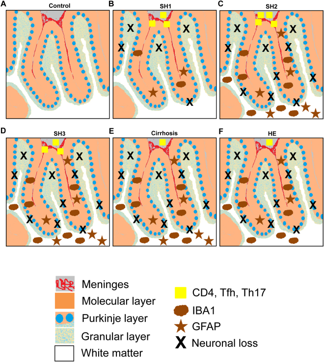
Figure 5.
Scheme summarizing the histo-architectural changes in cerebellum of patients with different grades of liver disease. (A) Cerebellum of a control subject with normal liver. (B) Patients at early stage (SH1) of steatohepatitis. An infiltration of CD4+ T lymphocytes (yellow squares) is observed in meninges (in red). CD4+ cells activate microglia (brown clouds) in the molecular layer (orange layer) and alter Bergmann glial fiber morphology (brown stars). Microglia and astrocytes are not activated in white matter (white color). Activated microglia in the molecular layer induces neurodegeneration (black X) of Purkinje (blue circles) and granular neurons (light blue spotted layer). (C) Patients at SH2 stage. The changes observed in SH1 are exacerbated, with increased microglia and astrocyte activation in molecular layer (orange). Microglia (brown clouds) and astrocytes (brown stars) are also activated in white matter (in white) and neuronal loss (black X) increases in Purkinje layer (blue circles). (D) Patients at SH3 stage. The changes observed are similar to SH2 patients. The only difference is a small decrease in CD4+ T lymphocytes (yellow squares). (E and F) Patients with liver cirrhosis and HE. A decrease of CD4+ lymphocytes (yellow square) infiltration is observed. Chronic neuroinflammation increases neuronal loss (black X) in Purkinje layer (blue circles). A significant reduction of GFAP content in white matter of patients with HE is observed (brown stars).

