
Figure 6.
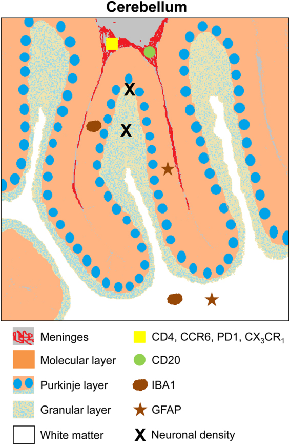
Scheme summarizing the analysis performed and their localization in cerebellum. Histo-architectural analysis was performed on post-mortem human cerebellum sections. Neuronal density analysis (black X) was performed in Purkinje layer (blue circles) and in granular layer (light blue spotted layer). Microglial (brown cloud, Iba1) and astroglial (brown star, GFAP) activation analysis was performed in molecular layer (in orange) and white matter (white). Infiltration of B lymphocytes (green circle, CD20) and of total T lymphocytes (yellow square, CD4) and the T lymphocytes subtypes Tfh, Th17 and CD4+CD28− was analyzed in meninges (in red).